Summary: Pigmentation disorders are conditions in which areas of skin become darker (hyperpigmentation) or lighter (hypopigmentation) than the surrounding skin. In Indian skin the commonest are melasma, post-inflammatory hyperpigmentation, solar lentigines, periorbital hyperpigmentation and lichen planus pigmentosus — driven by ultraviolet and visible light, heat, hormones, inflammation and genetics. They are highly treatable, but they are also easy to worsen: the wrong product or an overaggressive procedure can leave darker skin than it started with. Accurate diagnosis before treatment is the single most important step.
Dark patches, uneven tone, spots that will not fade — pigmentation is one of the most common skin concerns, and one of the most commonly mistreated.
The frustrating part is that pigmentation is easy to worsen. Getting it right requires knowing which type you have and how deep it sits, which is exactly where a dermatologist’s assessment changes the outcome.
This guide sets out the main pigmentation disorders, what causes them, and how they are treated safely in Indian skin.
What Are Pigmentation Disorders?
Pigmentation disorders are conditions in which melanin — the pigment that determines skin colour — is produced, distributed or retained unevenly.
Hyperpigmentation, the darkening type, is by far the commoner presentation and the usual reason patients seek treatment. Hypopigmentation, in which areas lose pigment, is less common but medically important.
The Biology, Briefly
Melanin is produced by melanocytes in the basal layer of the epidermis, packaged into organelles called melanosomes, and transferred to surrounding keratinocytes, where it forms a protective cap over the cell nucleus.
A point that surprises most patients: the number of melanocytes is broadly similar across all human skin. What differs between skin tones is the size, number, distribution and degree of degradation of the melanosomes, and the ratio of dark brown-black eumelanin to red-yellow pheomelanin.
Two further points explain most clinical behaviour. First, tyrosinase is the rate-limiting enzyme in melanin synthesis, which is why almost every depigmenting agent — and, unfortunately, mercury in illegal fairness creams — targets it. Second, once pigment escapes into the dermis (through an injured basement membrane, or engulfed by macrophages as melanophages) it clears far more slowly than epidermal pigment. That distinction between epidermal and dermal pigment is what determines how long treatment takes and how completely it works.
Indian skin, largely Fitzpatrick types IV and V, is particularly prone to pigmentation disorders. Higher constitutive melanin gives real photoprotective advantage, but it also means melanocytes respond more readily to any insult — which is why post-inflammatory hyperpigmentation is such a dominant problem here and why pigmentation is one of the most frequent reasons to see a dermatologist in India.
The main types of pigmentation disorder
Several conditions look broadly similar to the untrained eye and behave very differently. This is the single most useful section of this article.
| Condition | How It Presents | Key Point |
|---|---|---|
| Melasma | Symmetrical brown to greyish patches on cheeks, forehead, upper lip and nose | Chronic and relapsing; heat-sensitive; managed, not cured |
| Post-inflammatory hyperpigmentation (PIH) | Dark marks following acne, eczema, injury, waxing or an aggressive procedure | Follows the shape of the original lesion; usually fades, but slowly |
| Solar lentigines and ephelides | Discrete brown spots on sun-exposed areas; freckles | Among the most responsive to laser |
| Periorbital hyperpigmentation | “Dark circles” — often a mix of pigment, vascularity, thin skin and tear-trough shadow | Frequently not pigment at all; treating it as pigment fails |
| Lichen planus pigmentosus | Slate-grey to brown-black patches, often face, neck and flexures | Inflammatory and dermal; needs medical treatment, not lightening creams |
| Pigmented contact dermatitis (Riehl’s melanosis) | Diffuse brownish pigmentation, often from cosmetics, fragrances or hair dye | Identifying and removing the trigger is the treatment |
| Acanthosis nigricans | Velvety dark thickening of neck, axillae and knuckles | Often a marker of insulin resistance or obesity — warrants metabolic assessment |
| Hori’s naevus (ABNOM) and naevus of Ota | Bluish-grey dermal pigmentation, commonly over cheekbones | Routinely misdiagnosed as melasma; responds to laser, not to creams |
| Drug-induced pigmentation | Diffuse or patchy darkening linked to a medication | Needs drug history, not a peel |
| Hypopigmentation | Vitiligo, pityriasis alba, post-inflammatory hypopigmentation, idiopathic guttate hypomelanosis | Lighter, not darker — entirely different treatment pathway |
Hori’s naevus deserves particular mention. It is bilateral, sits over the cheekbones, and is very commonly treated as melasma for years without improvement — because it is dermal pigment that topical agents cannot reach. Distinguishing the two changes the entire plan.
Depth matters as much as diagnosis
Pigmentation is also classified by depth: epidermal (superficial, responds best), dermal (deeper, slower and less complete), and mixed (the commonest pattern in Indian skin). Wood’s lamp examination and dermoscopy help establish depth and pattern before treatment is chosen. This is not a formality — it is what prevents a patient being given three months of a cream that was never going to reach the pigment.
What causes pigmentation?
Sun is the dominant factor, but it is not the only one, and two important triggers are routinely left out of patient information.
Ultraviolet radiation: the leading trigger, worsening nearly all pigmentation, including brief daily exposure such as a commute.
Visible light, particularly high-energy blue light: induces pigmentation in darker skin types even without UV. Ordinary daylight and screens contribute. Most clear chemical sunscreens do not block it — which is why tinted formulations containing iron oxides matter in Indian skin.
Infrared heat: cooking over a flame, sun-facing travel and a humid coastal climate stimulate pigment independently of light. Hormonal changes: pregnancy, combined oral contraceptives, hormonal IUDs and thyroid dysfunction.
Inflammation: acne, eczema, injury, insect bites, waxing and threading — and over-aggressive cosmetic procedures.
Friction and pressure: a common and under-recognised cause on the neck, sides of the face and body.
Cosmetics, fragrances and hair dye: can cause pigmented contact dermatitis.
Genetics: a strong family tendency, particularly in melasma.
Medications: some antibiotics, antimalarials, antiepileptics, chemotherapy agents and others.
Steroid and fairness cream misuse: a leading iatrogenic cause of pigmentation in India, discussed below.
What to be careful of
Over-the-counter fairness creams. Independent testing has repeatedly found undeclared mercury and potent corticosteroids in such products. Mercury inhibits tyrosinase — which is precisely why it is added — and causes rashes, paradoxical discolouration, and nervous system and kidney damage. Prolonged facial steroid use causes topical steroid damaged face: atrophy, telangiectasia, acneiform eruption and a rebound flare on withdrawal, often with a superimposed steroid-modified fungal infection. If a cream lightens skin within days, that is a reason for suspicion, not reassurance.
Not every dark spot is “pigmentation.” A single lesion that is changing in size, shape or colour, has irregular borders, bleeds, itches persistently or looks different from your other marks needs dermatological assessment rather than a lightening cream. Sun spots and melanoma are not always distinguishable by eye, and dermoscopy exists for this reason.
How dermatologists treat pigmentation
Treatment is layered and built on photoprotection. Procedures sit near the top of that structure, not at the bottom, and are chosen for skin type rather than for novelty.
| Tier | What It Involves | Notes |
|---|---|---|
| 1. Photoprotection | Broad-spectrum SPF 50+, tinted with iron oxides to absorb visible light; reapplication; hats, shade, heat avoidance | Non-negotiable. Without it, every other treatment underperforms and relapse is near-certain |
| 2. Topical Therapy | Hydroquinone or modified Kligman-type combinations in defined cycles; azelaic acid, kojic acid, alpha arbutin, cysteamine, niacinamide, topical tranexamic acid, retinoids, vitamin C | Prescription agents, used for defined durations rather than indefinitely |
| 3. Treat the Driver | Control the acne, eczema or dermatitis; remove the offending cosmetic; investigate metabolic or thyroid causes; review medications | PIH will keep recurring until the underlying condition is treated |
| 4. Systemic Therapy | Oral tranexamic acid in selected melasma patients, after screening for thrombotic risk | Dermatologist-supervised only |
| 5. Procedures | Superficial chemical peels; low-fluence picosecond laser (PicoWay) | Adjunct, added once tiers 1–3 are established |
| 6. Maintenance | Ongoing photoprotection and periodic topical therapy | Most pigmentation conditions are chronic or relapse-prone |
Two cautions patients rarely hear. Hydroquinone is a prescription agent and prolonged uninterrupted use carries a risk of exogenous ochronosis — a paradoxical blue-grey pigmentation considerably harder to treat than the problem it was prescribed for. It belongs in supervised cycles with rest periods. And any peel or active used too aggressively on Fitzpatrick IV–V skin can itself trigger postinflammatory hyperpigmentation, producing exactly the outcome the patient came to avoid.
Where laser fits, and why the mechanism matters
Laser is an adjunct in pigmentation management, not a starting point. In darker skin the choice of device and settings is a safety decision, because heat and inflammation are themselves pigmentary triggers: a laser that clears pigment while depositing thermal energy into surrounding tissue can lighten a patch and provoke post-inflammatory hyperpigmentation or rebound within weeks.
The science: photoacoustic, not photothermal
Older Q-switched lasers deliver energy in nanosecond pulses — billionths of a second. That is long enough for a meaningful proportion of the absorbed energy to degrade into heat and conduct outwards into surrounding tissue. Pigment is destroyed largely photothermally: in effect, it is burned.
The Candela PicoWay delivers energy in picosecond pulses — trillionths of a second, roughly a thousand times shorter. The pulse ends faster than the melanosome’s thermal relaxation time, so the absorbed energy has no opportunity to disperse as heat. Instead it generates a rapid pressure wave that fragments the pigment particle mechanically. This is the photoacoustic, or photomechanical, effect.
The clinical consequences follow directly:
Pigment is broken into finer particles, cleared more efficiently by macrophages and the lymphatics.
Far less thermal energy reaches surrounding tissue, so the principal pigmentary trigger is largely avoided.
The risk of post-inflammatory hyperpigmentation — the characteristic hazard of laser in Indian skin — is meaningfully reduced.
Multiple wavelengths (1064 nm, 785 nm, 532 nm) allow target depth and pigment type to be matched, rather than treating everything on one setting.
PicoWay is not a heat-free device, and no honest clinic will claim otherwise. But because it works predominantly photoacoustically rather than photothermally, it permits a genuinely low-thermal-load approach in precisely the skin types where thermal load causes the most trouble.
PicoWay in Thane
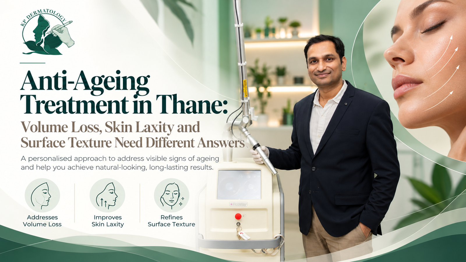
KP Dermatology is the only clinic in Thane equipped with the Candela PicoWay laser. Dr Prratyush More was the first dermatologist in Thane to introduce the technology, and every PicoWay procedure is performed by Dr More himself — not delegated to a technician or assistant.
Different pigmentation types call for different treatment. Solar lentigines respond briskly. Dermal pigment such as Hori’s naevus responds well but needs several sessions. Melasma is treated with deliberate restraint at low fluence, and only after photoprotection and topical therapy are established. Matching the approach to the diagnosis is the whole job.
Can pigmentation be treated permanently?
This depends entirely on which pigmentation you have, and the honest answer differs by condition — which is why a single blanket promise should make you cautious.
Solar lentigines and freckles can often be cleared substantially and durably, though new ones form with continued sun exposure.
Post-inflammatory hyperpigmentation generally resolves once the underlying inflammation is controlled — epidermal PIH over weeks to months, dermal PIH considerably longer.
It recurs if the driver recurs. Melasma is chronic. It is managed rather than cured, and relapses when photoprotection or maintenance stops.
Dermal naevoid pigmentation such as Hori’s naevus can clear well with laser and tends to stay clear.
Acanthosis nigricans improves when the underlying metabolic driver is addressed — creams alone achieve little.
Across all of them, most pigmentation improves noticeably over weeks to months rather than days, and results are held by maintenance rather than by the last session. Consistency beats intensity.
Frequently asked questions
How do I know which type of pigmentation I have?
Distribution, symmetry, colour, borders and history usually distinguish them, supported by Wood’s lamp and dermoscopy. This matters because melasma, PIH and Hori’s naevus can look similar and are treated quite differently.
How long does pigmentation treatment take?
Expect gradual improvement over weeks to months. Topical therapy typically shows visible change by eight to twelve weeks; laser results build across sessions. Anything promising clearance in days is not describing a treatment that respects the biology.
Will sunscreen alone clear my pigmentation?
It will not clear established pigmentation, but no treatment works reliably without it. Broad-spectrum SPF 50+, tinted to block visible light, both prevents relapse and improves the results of everything else.
Can laser make pigmentation worse?
Yes, if the device or settings are wrong for the skin type or diagnosis. Heat is a pigmentary trigger, so high-fluence treatment in Fitzpatrick IV–V frequently causes rebound. Low-fluence picosecond protocols, which act photo acoustically rather than photothermally, are considerably safer and are used as an adjunct.
Are home remedies and lemon juice safe?
Lemon juice is a common cause of phytophotodermatitis — a light-triggered reaction that leaves pigmentation worse than before. Abrasive scrubs and undiluted acids likewise cause inflammation, and inflammation is what produces PIH in the first place.
Does diet or a vitamin deficiency cause pigmentation?
Occasionally. Vitamin B12 deficiency can cause diffuse or acral hyperpigmentation, and thyroid dysfunction is associated with melasma. These are worth testing when the pattern suggests it — but diet is not the main driver of ordinary facial pigmentation.
Treating pigmentation safely with expert care
Because pigmentation is so easy to worsen, it needs a dermatologist willing to hold back as readily as intervene.
At KP Dermatology in Thane, the type and depth of pigmentation are diagnosed before anything is prescribed. Treatment is then built in layers — photoprotection, topical therapy, control of the underlying driver, and carefully selected procedures suited to Indian skin — with honest guidance on realistic results and the need for maintenance, rather than harsh one-size treatment. Dr Prratyush More brings over fourteen years of clinical experience and advanced international laser training, was the first dermatologist in Thane to introduce Candela PicoWay, and the clinic remains the only one in Thane with the technology.
Book a consultation
Pigmentation disorders are common and often stubborn, but with accurate diagnosis, appropriate treatment and diligent photoprotection, real improvement is very achievable. The key is treating your specific type correctly rather than guessing with products. If uneven tone or dark patches are bothering you, book a consultation at KP Dermatology, Thane, for a safe, personalised pigmentation plan.
References
1. Indian Pigmentary Expert Group. Consensus recommendations on the management of melasma in Indian patients.
2. Sarkar R, et al. Melasma in Indian patients: a multicentre clinico-epidemiological study. Indian Journal of Dermatology, Venereology and Leprology.
3. Davis EC, Callender VD. Postinflammatory hyperpigmentation: a review of the epidemiology, clinical features and treatment options in skin of colour. Journal of Clinical and Aesthetic Dermatology.
4. Mahmoud BH, et al. Impact of long-wavelength visible light on melanocompetent skin. Journal of Investigative Dermatology.
5. World Health Organization. Mercury in skin lightening products — information sheet.
6. Coondoo A, Phiske M, Verma S, Lahiri K. Side-effects of topical steroids: a long overdue revisit. Indian Dermatology Online Journal.
7. Anderson RR, Parrish JA. Selective photothermolysis: precise microsurgery by selective absorption of pulsed radiation. Science.
8. Torbeck RL, Schilling L, Khorasani H, et al. Evolution of the picosecond laser: a review of literature. Dermatologic Surgery.
This article is intended for general education and does not constitute individual medical advice. Pigmentation requires assessment by a qualified dermatologist before any treatment is started, and any single changing or unusual pigmented lesion should be examined promptly. Individual results vary and no outcome is guaranteed. Prescription topical and oral therapies mentioned here should be used only under dermatological supervision. Do not stop a long-term topical steroid without medical advice.